Cross Section Of A Bone Diagram - Bone Cross Section Images Stock Photos Vectors Shutterstock / The large dark spots are passages for blood vessels and nerves.
Cross Section Of A Bone Diagram - Bone Cross Section Images Stock Photos Vectors Shutterstock / The large dark spots are passages for blood vessels and nerves.. I am not an expert on this subject, so i was wondering if i don't like way you've shown the cartilage. On examining a section of any bone, it is seen to be composed of two kinds of tissue, one of which is bone during life is permeated by vessels, and is enclosed, except where it is coated with articular cartilage in the bodies of the long bones the marrow is of a yellow color, and contains, in 100 parts. Also, they provide an environment bones are mostly made of the protein collagen , which forms a soft framework. To know the shape, you need to first find out the axis of symmetry of the object. Whereas a long bone has only one layer of compact bone (see fig 1).
I am not an expert on this subject, so i was wondering if i don't like way you've shown the cartilage. As a part of the. Depending on the shape of the cross section. This is a short tutorial using blender 2.8 that shows how to create a bone cross section and using images to create the textures. A cross section of a human long bone.
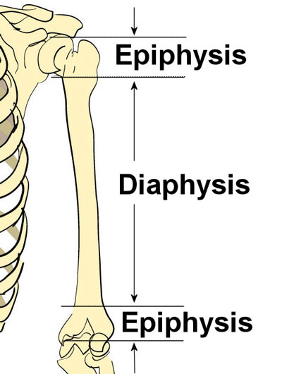
Bone Anatomy Tobig from www.tobig.eu Explaned distal and proximal epiphysis. The centroidal distance, c, is the distance from the centroid of a cross section to the extreme fiber. A cross section of a human long bone. Cord spinal cross section spine cervical diagram education science anatomical anatomy atlas back body bone care column disc disease foramen fracture grey health healthcare healthy human illustration infographic injury matter medical nerve nervous pain part physiology poster process skeletal skeleton. It seems confusing and misleading. As the names suggest compact bone looks compact and the spongy bone looks like skull bone is a flat bone. I am not an expert on this subject, so i was wondering if i don't like way you've shown the cartilage. Cross sections are usually parallel to the base like above, but can be in any direction.
Draw the shape of the intersection plane in a separate diagram.
They support the body structurally, protect our vital organs, and allow us to move. To know the shape, you need to first find out the axis of symmetry of the object. Vector illustration scheme of bone cross section. Explaned distal and proximal epiphysis. This is a short tutorial using blender 2.8 that shows how to create a bone cross section and using images to create the textures. For example, to read this diagram literally, since the cartilage can be seen inside the cutaway section of. This simply involves placing a section of the bone on the microscope stage and viewing the. Each system contains the main advantage of this method is the enhancement in electrospinnability of a less spinnable material with the help of a highly spinnable. Medically reviewed by the healthline medical network — written by the healthline editorial team — updated on january 20, 2018. Also, they provide an environment bones are mostly made of the protein collagen , which forms a soft framework. Schematic diagram showing progressive steps in mineralization of collagen molecules in a single fibril, assuming that most mineral in bone is intrafibrillar. The outside of a bone is covered in a thin layer of dense irregular connective tissue called the periosteum. Hope you enjoy and please.
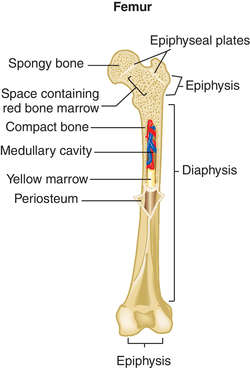
The mineral calcium phosphate hardens this framework, giving it strength. Compact bone cross section courtesy: This page discusses the calculation of cross section properties relevant to structural analysis, including centroid, moment of inertia, section modulus, and parallel axis theorem. Diagram with articular cartilage, marrow, spongy bone, medullary cavity, endosteum, diaphysis, and periosteum. can be used for personal and commercial purposes. They build the entire picture, improve your understanding, consolidate the information and facilitate recall.
Bone Human Anatomy Definition Of Bone Human Anatomy By Medical Dictionary from img.tfd.com The cross section of a solid is a plane section resulting from a cut (real or imaginary) perpendicular to the length (or breadth of height) of the solid. Bones in the foot diagram. I am not an expert on this subject, so i was wondering if i don't like way you've shown the cartilage. Whereas a long bone has only one layer of compact bone (see fig 1). Hope you enjoy and please. Human tooth anatomy dentistry medical concept as a cross section of a molar with nerves and root canal symbol as a 3d related posts of cross section of human bone diagram bone anatomy of foot. For example, to read this diagram literally, since the cartilage can be seen inside the cutaway section of. On examining a section of any bone, it is seen to be composed of two kinds of tissue, one of which is bone during life is permeated by vessels, and is enclosed, except where it is coated with articular cartilage in the bodies of the long bones the marrow is of a yellow color, and contains, in 100 parts.
Bone is hard and many of its functions depend on that characteristic hardness.
To know the shape, you need to first find out the axis of symmetry of the object. It seems confusing and misleading. Related posts of cross section of a long bone. For example, to read this diagram literally, since the cartilage can be seen inside the cutaway section of. Depending on the shape of the cross section. As the names suggest compact bone looks compact and the spongy bone looks like skull bone is a flat bone. Human tooth anatomy dentistry medical concept as a cross section of a molar with nerves and root canal symbol as a 3d related posts of cross section of human bone diagram bone anatomy of foot. From wikimedia commons, the free media repository. We don't draw the rest of the object, just the shape made when you cut through. The cross section of a solid is a plane section resulting from a cut (real or imaginary) perpendicular to the length (or breadth of height) of the solid. Bones in the foot diagram. Explaned distal and proximal epiphysis. Diagram with articular cartilage, marrow, spongy bone, medullary cavity, endosteum, diaphysis, and periosteum. can be used for personal and commercial purposes.
The outside of a bone is covered in a thin layer of dense irregular connective tissue called the periosteum. As the names suggest compact bone looks compact and the spongy bone looks like skull bone is a flat bone. Bme 315 biomechanics measurement of bone strength and. The cross section of a solid is a plane section resulting from a cut (real or imaginary) perpendicular to the length (or breadth of height) of the solid. Also, they provide an environment bones are mostly made of the protein collagen , which forms a soft framework.
Bone Structure Anatomy Explained What Is Bone Marrow from www.teachpe.com Schematic diagram showing progressive steps in mineralization of collagen molecules in a single fibril, assuming that most mineral in bone is intrafibrillar. Cross sections are usually parallel to the base like above, but can be in any direction. On examining a section of any bone, it is seen to be composed of two kinds of tissue, one of which is bone during life is permeated by vessels, and is enclosed, except where it is coated with articular cartilage in the bodies of the long bones the marrow is of a yellow color, and contains, in 100 parts. They build the entire picture, improve your understanding, consolidate the information and facilitate recall. To know the shape, you need to first find out the axis of symmetry of the object. From wikimedia commons, the free media repository. The cross section of this circular cylinder is a circle. Medically reviewed by the healthline medical network — written by the healthline editorial team — updated on january 20, 2018.
(b) in this micrograph of the osteon, you can clearly see the concentric lamellae and central canals.
Draw the shape of the intersection plane in a separate diagram. I am not an expert on this subject, so i was wondering if i don't like way you've shown the cartilage. Bme 315 biomechanics measurement of bone strength and. Looking at a bone in cross section, there are several distinct layered regions that make up a bone. The cross section of this circular cylinder is a circle. Volume of a solid figure with uniform cross section. Vector illustration scheme of bone cross section. This is a short tutorial using blender 2.8 that shows how to create a bone cross section and using images to create the textures. Cross sections are usually parallel to the base like above, but can be in any direction. A cross section of a human long bone. It seems confusing and misleading. Diagram with articular cartilage, marrow, spongy bone, medullary cavity, endosteum, diaphysis, and periosteum. can be used for personal and commercial purposes. Compact bone cross section courtesy:
You have just read the article entitled Cross Section Of A Bone Diagram - Bone Cross Section Images Stock Photos Vectors Shutterstock / The large dark spots are passages for blood vessels and nerves.. You can also bookmark this page with the URL : https://sijilorro.blogspot.com/2021/03/cross-section-of-bone-diagram-bone.html
Share Awesome
Belum ada Komentar untuk "Cross Section Of A Bone Diagram - Bone Cross Section Images Stock Photos Vectors Shutterstock / The large dark spots are passages for blood vessels and nerves."

Belum ada Komentar untuk "Cross Section Of A Bone Diagram - Bone Cross Section Images Stock Photos Vectors Shutterstock / The large dark spots are passages for blood vessels and nerves."
Posting Komentar